Protoplast fusion
and genetic analysis
in Cephalosporium acremonium

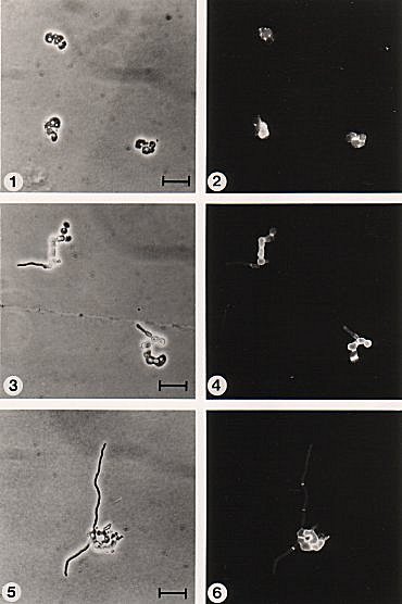
Figure 4.1 Regeneration and reversion of protoplasts of C. acremonium on SMS observed using phase contrast and fluorescent microscopy illustrating the typical sequence of events. (1) and (2) After 16 h incubation; small patches of fluorescence were clearly visible on the regenerating protoplasts. (3) and (4) After 32 h; outgrowth of short chains of brightly fluorescing "yeast-like" cells. (5) and (6) After 40 h; climax of protoplast reversion with the development of normal hyphae. The bar markers represent 20 µm. |
Copyright © 1982 Paul F Hamlyn
(http://fungus.org.uk/cv/thesis_fig4.1.htm)