Protoplast fusion
and genetic analysis
in Cephalosporium acremonium
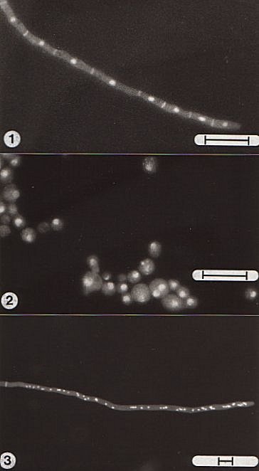
3.2.6 Cytological examinations Nuclear staining was carried out by the method of Slater (1976) with the following modifications. Chromomycin A3 (Sigma) was used instead of mithramycin and it was found necessary to fix protoplast preparations before staining. Protoplasts were fixed in 4% (v/v) glutaraldehyde in 0.8 M mannitol for 1.5h at room temperature. The fixative was removed by centrifugation and the pellet resuspended in distilled water. An equal volume of the stain (0.4 mg ml-1 chromomycin in 50% aqueous ethanol containing 30 mM MgCl2) was then added and the material examined as a wet mount. Whole mycelium was also stained to investigate the distribution of nuclei in hyphae. No fixation was required and washed mycelium was mounted in the stain. To reveal septa the preparations were counterstained with 0.0005% (v/v) aqueous Tinopal (Ciba-Geigy). All the samples were examined under a Vickers M17 uv-fluorescence microscope containing the following filters: Exciter: BG 3 (5 mm) Dichroic: Violet Barrier: G 475 (4 mm) Photographs were taken using Ilford HP5 (400 ASA) film. Storage of the stained material at 4oC for 24h improved the staining. The hyphal compartments of C. acremonium are uninucleate and the majority of protoplasts were also found to be uninucleate. In contrast, protoplasts isolated from mycelium of Penicillium chrysogenum or Aspergillus nidulans are often multinucleate reflecting the multinucleate nature of hyphal cells in these organisms (Fig. 3.11).
Reference Slater, M. L. (1976). Rapid nuclear staining method for Saccharomyces cerevisiae. Journal of Bacteriology, 126, 1339-1341. |
Figure 3.11 Fluorescent staining of nuclei in hyphal cells and protoplasts of Cephalosporium acremonium. A hypha of Aspergillus nidulans is included for comparison. (1) Hypha of C. acremonium stained with Chromomycin and Tinopal, showing nuclei and septa. The uninucleate nature of the hyphal compartments in this organism are clearly illustrated. (2) Protoplasts of C. acremonium stained with Chromomycin. The protoplasts were fixed in 0.8 M mannitol containing 4% glutaraldehyde prior to staining. Note the rare binucleate protoplast. (3) Hypha of A. nidulans stained with Chromomycin and Tinopal. Note the multinucleate hyphal tip. The bar markers represent 10 µm. |
Copyright © 1982 Paul F Hamlyn
(http://fungus.org.uk/cv/thesis_3.2.6.htm)